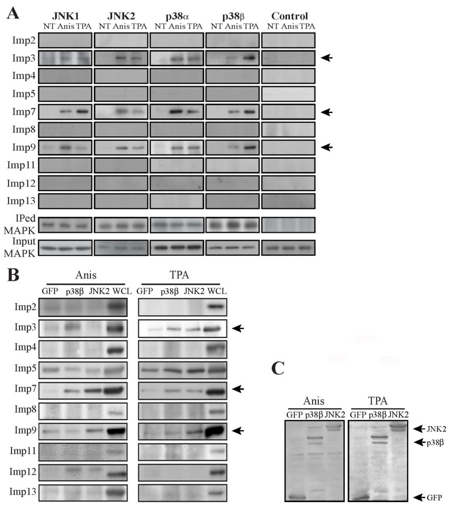
Fig. 5. Endogenous and overexpressed JNKs and p38s CoIP with Imps 3, 7 and 9. (A) A CoIP screen to detect the Imps interacting with JNK1/2 and p38α/β in MCF7 cells. MCF7 cells were grown to 70% confluence, serums starved and then were stimulated with either anisomycin (Anis 0.5 μg/ml, 15 min), TPA (250 nM, 15 min) or left untreated (NT). Cells extracts were then subjected to CoIP with the indicated Abs. Imps, IPed MAPKs and inputs (in extracts) were detected by Western blot with the indicated Abs. (B) Interaction of overexpressed MAPKs with Imps 3,7 and 9 in HeLa cells. HeLa cells were transfected with either JNK2-GFP (wt-JNK2), p38β-GFP (wt-p38β) or GFP alone. Cells were serum starved, and then treated as described and subjected to CoIP with anti GFP Abs. The interacting Imps and the loading extracts were detected using Western blot analysis with the indicated Abs. Arrows indicate the interacting Imps. WCL is whole cell extract. (C) Loading control of overexpressed JNK2 and p38α. Extracts of HeLa cells overexpressing either JNK2-GFP or p38α-GFP or GFP alone were subjected to Western blot analysis with anti GFP Abs. The experiments were reproduced twice. The antibodies detected mainly the two bands (two start-sites) of the GFP-MAPKs and also few minor degradation products/impurities.